

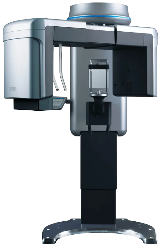
Cone Beam Computed Tomography (CBCT)
CBCT stands for Cone Beam Computed Tomography. It's a 3D imaging technology used in dentistry to provide detailed images of the oral and maxillofacial regions. CBCT scans are crucial for precise treatment planning, especially for implant placement, oral surgery, and orthodontics.
Dental practitioners can benefit from a cone beam in various ways. These consist of:
Evaluation of Endodontics
Orthodontic-related impacted teet
assessment of the paranasal sinuses
Visualizing the TMJ
trauma assessment
immediate advanced imaging and medical care
However, One of the most common reasons dental professionals seek cone beam technology is to offer dental implants and other specialized treatments to their patients. Cone beam computed tomography (CBCT) lets you visualize a cross-sectional view of the implant location. This view provides critical information you can't get from a 2D image. The AAOMR has been recommending cross-sectional imaging for assessing all dental implant sites since 2012, and cone beam is the method of choice.
Explore More!


Software Features
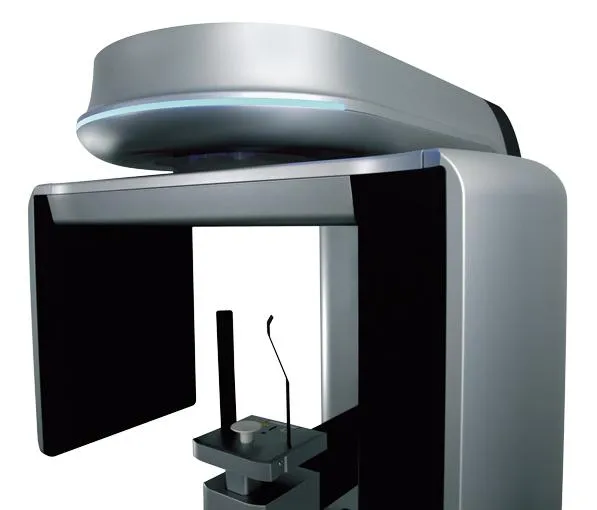
With the precision and professional competence of CBCT, dental professionals have a powerful partner at their side. The powerful system components of this model enable an extraordinary combination of the most precise 3D dental imaging, large image detail, low radiation exposure, reliable diagnostics and digital planning. Its patient management system is designed for secure and networked communication of patient data across multiple rooms within a practice and can be integrated into the existing infrastructure with ease. Meet the world’s first 3D CBCT with integrated ceph X-ray. With its superior resolution, unprecedented versatility and compact size, it is a game changer in dental imaging.
Multi-data
Load multiple patient scans on a single screen. Synchronize pre- and post-operative scans and detect differences slice by slice.
Patient Education and Presentation
Quickly capture 3D animated video clips for patient education and lecture presentations. Increase case acceptance through better patient understanding.
Collaborative Tools
Automatically save 3D image reports to an MS Word template and attach to the patient’s practice management record. Collaborate with referring dentists by burning a patient disc with the sample viewer. Capture and email images quickly.
Remote Access
Work on cases from home or a satellite office without long connectivity delays. Lead virtual treatment planning meetings remotely with PreXion3D.
Thin Client Server
PreXion3D CBCT scanners do not require computer hardware upgrades. Their software does not slow down network bandwidth like other CBCT scanners.
Implant Library
Use our extensive library or customize your own.
Save Scenes
Save your case workup as a scene or create multiple saved scenes with a single scan.
3D Templates
Save time with over 20 pre-made 3D volume rendering templates or customize your own.
Slab and Cutting
Slab feature allows the clinician to see inside structures while rotating the 3D image. Cut away structures to see exactly what is pertinent to your case.
Highlights:
Accurate 360-degree gantry rotation
260-1,024 projected views
Dedicated 2D pan mode option
Clearest detail with a 0.3mm focal spot & 0.08-0.2mm voxel
Key Features:
Innovative built-in ceph arm design freeing over 2 feet of space
0.3×0.3mm focal spot – the smallest in the industry • Multiple fields of view (FOV) ranging from 5×5 to 15×16.
Low dose, HD and endo CT modes with scan times of 20 seconds or lower
14-second pano and 16-second ceph scan times
Clinical Applications:
Implant placement surgery
Endodontics
Periodontics
Orthodontics
Oral-maxillofacial surgery
TMJ treatment
Pathology
Impacted and supernumerary teeth
DICOM export for implant surgical guides & CAD/CAM integration
Airway analysis